Immune Checkpoint Inhibitor Targeting LAG3 May Lead to Therapeutic Approaches in Advanced Melanoma
Immune checkpoint inhibitors are a therapy that prevents tumors from shutting down the immune response, allowing T-cells to kill cancer cells. Checkpoint inhibitors have transformed cancer care, specifically in treating a variety of solid tumor types such as melanoma and lung cancer, by targeting the proteins PD-1 and CTLA-4. However, new advances have been made as the U.S. Food and Drug Administration recently approved a new immune checkpoint inhibitor targeting the protein LAG3. An anti-LAG3 antibody, called relatlimab, was administered in combination with the anti-PD-1 antibody nivolumab to treat advanced melanoma.
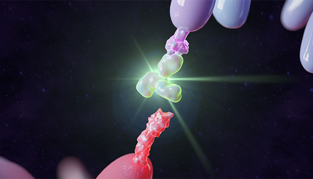
Little has been known about the structure of the LAG3 protein. In the absence of a three-dimensional structure, LAG3-based drugs must be designed “in the dark” using inefficient screening methods. A team of Moffitt researchers has become the first in the world to visualize the molecular structure of the LAG3 protein. Dr. Vince Luca, an assistant member of the Drug Discovery Department, presented their findings in an article published in Nature Immunology, describing the crystal structure of LAG3 and how it interacts with molecules produced by cancer cells.

"When I started my lab at Moffitt, I noticed a growing interest in LAG3 as an immunotherapy target," Dr. Luca said. "I was surprised at how little we knew about the LAG3 structure and its molecular mechanism, despite about 30 years of literature highlighting its role in the immune system.”
Dr. Luca and his team used X-ray crystallography to "see" the structure of the LAG3 protein at nearly atomic resolution. Moffitt’s researchers mapped out the regions of LAG3 that bound to signaling molecules FGL1 and MHCII and two different anti-LAG3 antibodies. Based on this information, the team was able to determine which antibody binding sites were ideal to inhibit LAG3 activity. They discovered how structural interactions of LAG3 and FGL1 inhibit T-cell function and found that the binding of these two molecules causes LAG3 to cluster on the surface of T-cells. The researchers theorize this may contribute to the inhibitory activity of LAG3 by blocking the T-cells from properly recognizing tumor cells.
The data reveal several important insights into the three-dimensional structure of LAG3 and how it interacts with other molecules, potentially leading to better targeted therapeutic approaches in the future.
"Collectively, our structural, epitope mapping and functional studies provide an improved framework for understanding LAG3 molecular function. In the future, additional structures of LAG3 bound to ligands and antibodies will refine our knowledge of the LAG3 signaling axis to illuminate how extracellular binding events fine-tune LAG3-mediated changes in T cell activity. In turn, such structural insights should guide the development of maximally effective LAG3-based immunotherapies," said Dr. Luca.
This study was supported by the National Institutes of Health (R35GM133482, P01AI120943, R01CA230610, P30CA076292), the V Foundation for Cancer Research and the Rita Allen Foundation Scholars Program.
If you’d like to refer a patient to Moffitt, complete our online form or contact a physician liaison for assistance. As part of our efforts to shorten referral times as much as possible, online referrals are typically responded to within 24 - 48 hours.