What is Spindle Cell Sarcoma?
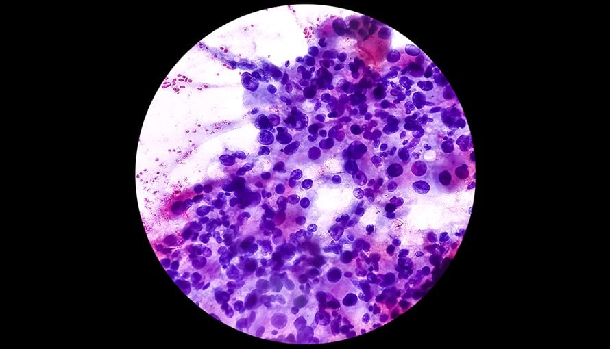
A spindle cell sarcoma is a soft tissue tumor that originates in a bone. When viewed under a microscope, the cancerous cells appear long and narrow (spindle-shaped). While this rare malignancy can affect anyone, it is most frequently diagnosed in adults 40 and older.
Most spindle cell sarcomas are found in a long bone, such as the thigh bone (femur), shin bone (tibia) or upper arm bone (humerus). Additionally, a subtype of spindle cell sarcoma known as undifferentiated sarcoma of the bone is often found in the pelvis.
What are the signs of spindle cell sarcoma?
Because a spindle cell sarcoma can develop in any bone, the site of the symptoms can vary. In general, they include:
- Bone pain, swelling and tenderness
- Fractures due to bone weakening
- A lump or mass that can be felt
- General malaise
- Extreme fatigue
What causes spindle cell sarcoma?
As part of the body's natural response to a soft tissue injury, spindle cells in the damaged tissue will begin dividing to promote healing. Normally, once the affected area is restored, the spindle cells will stop replicating. Sometimes, however, the cells may continue to divide uncontrollably for reasons that are not fully understood. The excess cells may then build up, bind together and form a spindle cell sarcoma.
What are the risk factors for spindle cell sarcoma?
Some people are genetically predisposed to spindle cell sarcoma. Other risk factors include:
- Paget’s disease of the bone – A chronic bone condition, Paget’s disease interferes with the bone remodeling process and causes the bones to become weak and misshapen.
- Prior radiation therapy to a bone – A spindle cell sarcoma may form many years after a bone was exposed to radiation therapy.
- Fibrous dysplasia – As part of the bone remodeling process, mature bone tissue is replaced with undeveloped connective tissue instead of new bone tissue.
- Bone infarction – Due to a poor blood supply, bone tissue is deprived of essential oxygen and dies.
- Osteomyelitis – Often a result of an injury, osteomyelitis is a bone infection that causes inflammation and swelling.
How is spindle cell sarcoma diagnosed?
If a spindle cell sarcoma is suspected, a physician may order one or more imaging tests, such as an:
- X-ray – A small dose of ionizing radiation is used to create pictures of a bone.
- Computerized tomography (CT) scan – Several X-rays are combined by a computer to create three-dimensional images of a bone.
- Magnetic resonance imaging (MRI) scan – Strong magnetic fields and radio waves are used to produce highly detailed images of a bone.
- Positron emission tomography (PET) scan – Images are captured after a radioactive tracer is administered. Because cancerous cells will absorb the radiotracer more rapidly than healthy cells, the sarcoma will be highlighted in the resulting images.
- Isotope bone scan – After a small amount of radioactive fluid (radionuclide) is injected, images are captured with a gamma camera, which can reveal “hot spots” where cancerous cells may be present.
- Bone biopsy – A small sample of a suspicious bone lesion is taken and examined under a microscope by a pathologist, who can identify cancerous cells.

#1 Cancer Hospital in Florida
Schedule an AppointmentWhat are the treatment options for spindle cell sarcoma?
Treatment for spindle cell sarcoma can vary based on the location and stage of the tumor. In many cases, treatment is done in phases and includes a combination of chemotherapy, surgery and radiation therapy.
- Chemotherapy – The first cycle of chemotherapy may be administered before surgery to help shrink the tumor and make it easier to remove. One option is an isolated limb infusion (ILI), which involves delivering a high dose of chemo directly to a sarcoma in an arm or leg. After surgery, chemotherapy may resume and continue for several more weeks.
- Surgery – The goal of surgical treatment is to safely remove as much of the tumor as possible while preserving normal bodily function. For instance, limb-sparing surgery may be performed to remove a sarcoma in an arm or leg without removing the entire limb.
- Radiation therapy – After surgery, radiation therapy may be administered to target residual cancer cells and help prevent a recurrence. It may also be used as a standalone treatment for a sarcoma that cannot be surgically removed due to its location.
Sarcoma treatment at Moffitt Cancer Center
At Moffitt, cancer care is directly tied to research, and we continue to explore promising new treatment options for all types of sarcomas, including spindle cell sarcoma. We know that better science leads to innovative clinical trials that will one day become the next standard of care. In recognition of our groundbreaking achievements to date, the National Cancer Institute has awarded us the prestigious designation of Comprehensive Cancer Center.
If you would like to learn more about spindle cell sarcoma, contact Moffitt Cancer Center at 1-888-663-3488 or complete our new patient registration form online to request an appointment. You can consult with a specialist in our Sarcoma Program with or without a referral.