Ependymoma Overview
An ependymoma is a type of tumor that forms in the ependymal cells of the brain and spinal cord. Also known as radial glial cells, these specialized epithelial cells line the ventricular system of the brain and play a vital role in the production of cerebrospinal fluid (CSF). In addition to delivering essential nutrients to the brain and spinal cord, CSF cushions and protects these sensitive structures. Although ependymoma can travel to other areas of the central nervous system (CNS) via the CSF, the cancer rarely spreads to other areas of the body.
While ependymomas can develop at any age, these low-grade gliomas are most frequently diagnosed in young children.
Ependymoma causes and risk factors
Like all types of cancer, ependymoma results from harmful changes to the genes that control cellular function, allowing for rapid and uncontrolled cell growth. The precise triggers of the gene mutations that lead to the development of ependymoma are unknown.
Most ependymomas occur sporadically. However, researchers have discovered that children who have an inherited condition known as neurofibromatosis type 2 (NF2) have an increased risk of developing ependymomas and other CNS tumors.

Ependymoma signs and symptoms
The most common symptom of a brain tumor is a persistent or unusually severe headache. An ependymoma can also cause:
- Nausea and vomiting
- Blurred or double vision
- Loss of vision
- Impaired balance
- Dizziness
- Jerky eye movements
- Seizures
- Neck or back pain
- Numbness
- Muscle weakness
Additionally, a child with ependymoma may regress in development or reach developmental milestones more slowly than expected.
Ependymoma diagnosis
In addition to performing a physical examination, a physician may order one or more tests to diagnose ependymoma, such as a:
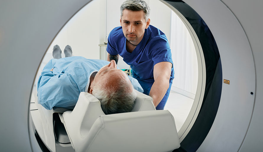
- Computed tomography (CT) scan - X-rays taken from multiple angles are combined by a computer to create a three-dimensional image.
- Magnetic resonance imaging (MRI) scan - Magnetic fields are used to produce highly detailed images.
- Lumbar puncture (spinal tap) - A needle is used to obtain a CSF sample for microscopic evaluation by a pathologist.
- Biopsy - A small amount of suspicious tissue is surgically removed for microscopic evaluation by a pathologist.
Although any of these diagnostic tests may suggest a brain or spinal cord tumor that warrants follow-up, only a biopsy can be used to definitively diagnose ependymoma.
Ependymoma treatment
Surgery is usually the preferred treatment for a suspected ependymoma. After temporarily removing a piece of skull bone to access the tumor site, a neurosurgeon will remove as much of the tumor as safely possible along with a slim margin of surrounding healthy tissue. In addition to providing a tissue sample to confirm the diagnosis, surgery can help relieve the symptoms caused by a tumor that is pressuring the brain or spinal cord. In general, the prognosis is best if the tumor can be completely removed (a total resection), which may involve more than one surgical session.
If an ependymoma is deemed inoperable (unresectable) due to its inaccessible location or proximity to vital structures, other treatment options may be considered, such as radiation therapy, chemotherapy and clinical trials.
Benefit from world-class care at Moffitt Cancer Center
As a proud member of the National Cancer Institute’s Adult Brain Tumor Consortium, Moffitt is committed to advancing research into the most effective diagnostic and treatment strategies for all types of brain and spinal cord tumors, including ependymomas. Our patients can benefit from the latest options in our renowned Neuro-Oncology Program, along with promising new therapies currently available only through our robust clinical trials portfolio.
If you would like to learn more about ependymoma, you do not need a referral to consult with an expert at Moffitt. Request an appointment by calling 1-888-663-3488 or submitting a new patient registration form online.
