Heavy Artillery in the Battle Against Brain Tumors
A lot of life’s biggest moments can happen in a short span of time. An unforgettable wedding. A jubilant baby announcement. A milestone birthday celebration. A lifechanging brain surgery.
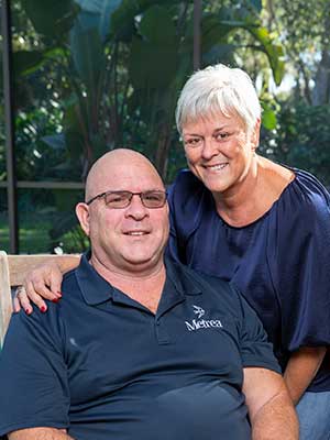
Robert Roggeman started out the summer of 2023 with a carefree calendar. He and his wife, Lisa, had traveled to a family friend’s wedding in Huntsville, Alabama, the first week of June. The celebration was beautiful. But on the flight back home to Tampa, the retired Army colonel experienced a jolting end to the trip. On the plane’s descent, he was hit with a splitting headache, the pain so intense it made his eyes water.
The one-time tank commander had traveled all over the world in the course of his military career, and he had experienced headaches on planes before. He was not one to rush to a doctor. But the next morning, Robert’s eye was red and swollen shut. Worried that he’d had a stroke, his wife insisted he get checked out.
“Usually I’d fight her because I don’t want to go to the doctor,” Robert said. This time, he reluctantly agreed.
Robert’s primary care doctor examined his eye and, at Lisa’s request, sent him for an MRI to check for signs of a stroke. He also referred Robert to an ophthalmologist. The eye doctor cleared Robert, but the MRI results showed something unexpected. Robert hadn’t had a stroke. Instead, there was a suspicious spot on his brain.
The couple and their daughters, Julie and Maggie, were nervous. The family had been through the stress of the unknown before with deployments. Julie, 29, feared the worst. Maggie, 27, reassured her dad everything would be fine.
Robert and Lisa made an appointment at Moffitt Cancer Center.
‘Very Fortuitous’
In July 2023, Robert and Lisa met with Michael Vogelbaum, MD, PhD, chief of neurosurgery and leader of the Neuro-Oncology Program at Moffitt. Vogelbaum explained that the MRI results had revealed a brain tumor called a glioma.
Robert and his wife, Lisa, happened to come to Moffitt the same month the new surgical hospital opened. He was the first patient to benefit from the new intraoperative MRI.
“He told us: ‘You guys are very fortuitous,’” Robert remembered. “‘No. 1, you listened to your wife. No. 2, you took some action. No. 3, the incident on the plane has nothing to do with the brain tumor.’”
It turns out the headache and the swollen eye were symptoms of unrelated sinus pressure. The brain tumor was not causing any symptoms yet. But the MRI had caught it early.
“Fortuitous” is a word that stuck with Robert. His father had died of a glioblastoma, an aggressive type of brain tumor, when he was 80. By the time his father had symptoms, it was too late to operate. Because Robert’s tumor was diagnosed early, his prognosis was much better.
In light of his family history, Robert opted to do genetic testing so he could better prepare his daughters if there was any risk for them down the line. He quickly learned his girls were fortunate — there was no genetic linkage.
The timing of Robert’s diagnosis was also serendipitous. In July, the cancer center opened its new state-of-the-art Moffitt McKinley Hospital, which houses an innovative intraoperative magnetic resonance imaging (iMRI) system. The iMRI allows doctors such as Vogelbaum to conduct real-time imaging during delicate brain surgeries without moving the patient. It improves the surgeon’s ability to remove as much of the cancer as possible.
Robert’s brain surgery was set for Aug. 8, 2023. He would be the first patient to benefit from the iMRI.
‘Millimeters Matter’
An internationally recognized leader in neurosurgical oncology, Vogelbaum has been using various forms of imaging technology in surgery for decades. He has experienced the evolution of the iMRI technology and been a champion for bringing the system to Moffitt, knowing it would improve outcomes for patients like Robert.
Traditionally, brain surgeons have been guided by imaging that is done preoperatively. Vogelbaum explains that in the early years of his career, a preoperative CT scan or MRI image would be displayed on a light board in the OR and used to estimate where on the head the opening should be made.
“And when you were removing the tumor, all you had were visual cues and an understanding of where the tumor had been in the anatomy,” he said.
A revolution occurred with computerized image guidance technology, which uses preoperative MRI scans and patient positioning data to help surgeons better pinpoint tumor locations. But that technology also has its limitations.

“The brain is not rigid,” Vogelbaum explained. “As time goes by and the brain relaxes while we are working on it, that image guidance becomes less and less accurate because it’s based on a preoperative MRI. And in our world, millimeters matter.”
That’s why the intraoperative MRI is a gamechanger. It allows surgeons to get real-time imaging on a patient while the person is still in the operating room and under anesthesia. If the scan shows that there is abnormal tissue remaining, the surgeon can continue to operate and remove it.
“The intraoperative MRI helps us to do the best we can in terms of removing the tumor so we can be more confident that we’ve accomplished that,” Vogelbaum explained. “There are situations where we cannot remove all of the tumor safely. But we want to eliminate the situations where we could have removed all of the tumor but didn’t.”
Happiness Among Hard Times
Robert and Lisa were impressed with Vogelbaum’s experience and expertise. They trusted that they were in good hands, but the weight of what was to come hung heavy over them.
With Robert’s surgery scheduled, Lisa immediately began rallying the troops to support the family. For years, she had led and been a part of Family Readiness groups that support service members and military families during deployment, relocations and unexpected emergencies. This time, she assembled a group of family members, friends and close neighbors as a support system.
“We know about grieving,” Robert said. “We were hoping for the best but preparing for the worst.”
Amid the shock, stress and sorrow, though, life’s bright moments continued to shine through.
The week before surgery, the couple got some uplifting news. Their younger daughter, Maggie, was pregnant. The baby would be their first grandchild.
On the Saturday before the surgery, the family celebrated another milestone — Robert’s 60th birthday. Lisa and the girls threw him an epic party at the historic Centro Asturiano in Ybor City. Relatives flew in from all over. Old military buddies rallied by his side. Robert was inundated with personalized mementoes and engulfed in decades of friendship.
The glow of the party carried Robert and Lisa through the weekend, old stories and new memories taking front stage.
By Monday, though, the family’s focus turned to surgery preparations. And on Tuesday morning, Robert was rolled into the operating room.
The Tough Part
Once Robert was taken back for surgery, Lisa sat on edge in the family waiting area, anxious for updates on her husband of 32 years. She knew Robert was tough, but waiting is never easy.
Vogelbaum’s initial work to remove the tumor took about an hour. The surgical team reported back to Lisa regularly to let her know that everything was going well.
Once Vogelbaum reached the point where he needed an updated image of the surgical area, the team prepared Robert for the iMRI. Vogelbaum made sure there was no active bleeding, filled the surgical cavity with saline and performed a temporary closure. The team then carefully covered Robert with sterile towels and a large plastic drape, essentially cocooning him to protect the sterile field.
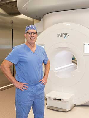
Vogelbaum and his team used the iMRI to get new images of Robert's brain during surgery, enabing the doctor to continue resecting the tumor.
Housed in a shielded diagnostic space between two operating rooms, the iMRI is a multiton device that hangs from a track on the ceiling. It can smoothly glide into the ORs on either side for intraoperative imaging. Before the massive magnet could enter the operating room, though, a host of safety checks needed to be performed.
Ferromagnetic materials and equipment had to be pushed outside of the safety zones marked on the operating room floor, indicating where they were safe from the iMRI’s powerful magnetic forces. Monitors, carts and other equipment were rolled away from Robert and out of the magnet’s reach. Surgical lights, which are mounted on large arms attached to the OR walls, were swung back to the edges of the room for safety. Robert himself had already been carefully checked to ensure there was no risk of burns from metallic implants.
With the patient and the operating room prepped, a specially trained safety officer ran through a final checklist to make sure everything was in place for the iMRI’s entrance. When the safety officer gave the all clear, the iMRI bay doors opened and the magnet moved into place, perfectly incapsulating Robert. The team performed a new scan, and then the iMRI receded back into its space.
“Most of the time, when I use the intraoperative MRI, I don’t end up removing additional tissue,” Vogelbaum explained. “However, the few times I do end up removing additional tissue, I’m grateful for the fact that I was able to discover that intraoperatively instead of postoperatively.”
In Robert’s case, the intraoperative imaging made a difference. With a new, more accurate view of the surgical area, Vogelbaum went back to work resecting the tumor.
All told, the surgery took three hours. When it was complete, Robert was rolled out of the operating room and into the intensive care unit to begin his recovery.
Back to Life
One of the first things Robert thought about when he woke up from brain surgery was lunch. The nurse gave him a menu and asked about his pain level. He estimated it at a 3.
“When I woke up, I didn’t have a lot of pain. I didn’t have a splitting headache like I thought I was going to have,” he said.
Vogelbaum had told Robert that most patients stay in the hospital for two nights after brain surgery, but Robert was ready to go home immediately.
“What do I have to do to get out of here?” he asked the nurse.
First, he needed a postoperative MRI to make sure everything was looking good. After one night in the ICU, though, he was cleared to go home.
The discharge was an emotional experience for the family. So much pent-up distress.
Robert reassured his girls: “I feel great. Don’t worry about me. You guys take care of Mom. I’ll be fine. It’s time to go home.”
Anticipating a rough recovery, Robert had taken the rest of the week off from his job as vice president of operations for a local defense company. Julie, who lives in New Smyrna Beach, was staying at the house to help. Maggie, who lives nearby in Tampa, was also at his side. Friends and family members were on standby to assist.
But Robert, with 24 staples in his head, didn’t feel like laying around. He was able to walk and get around normally. He wanted to go back to work.
“My family was flabbergasted,” he said.
He decided to work the rest of the week from home. The next week, he hadn’t been cleared to drive yet, so he got a ride into the office from a co-worker who lives nearby.
“To me, it’s amazing that I could recover so quick and get back to normal,” he said.
And there was more reason for optimism. When the pathology reports came back, Vogelbaum explained that the tumor was a grade 2 glioma. The outlook was positive.
“It was the best-case scenario,” Robert said. “They explained it’s not a real aggressive type of tumor. We think we got it all. And we will continue to monitor.”
Vogelbaum was also pleased with the outcome and has high expectations for the benefit the iMRI holds for future patients with brain tumors.
“We have ever-growing bodies of evidence showing that the more we can remove safely, the better the patient is likely to do in terms of maximizing survival,” he explained.
For Robert, ongoing monitoring requires a follow-up MRI every three months. He had his first in early November, and everything looked good.
“For me, this is a second chance or a continuation to live a good life,” he said, “to be healthy and try to do good.”
November marked five months since the fateful plane ride that led to Robert’s diagnosis. The family had packed in a lot of joy, tears, trepidation and hope in that relatively short amount of time.
Robert and Lisa were ready to move forward. The week after his first follow-up scan, the couple set sail on a weeklong cruise to the eastern Caribbean. In early December, the family traveled to Boston to catch the Army-Navy game. By Christmas, the incision scar across the front of Robert’s skull had faded to be almost invisible, a faint reminder of the family’s big battle.
With the whirlwind of the summer in the rearview, Robert and Lisa were looking forward to the new year. And of course life’s next major event: the birth of their first grandbaby.