16MB044 Molecular Imaging Probe: Novel IDO1-Targeting Cancer Diagnostic PET Imaging Agent
IDO1 (indoleamine 2,3-dioxygenase 1) is a tryptophan-metabolizing enzyme that mediates an immunosuppressive tumor environment whereby tumors can evade the immune system. High levels of IDO1 have been detected in a variety of tumors, and there are several IDO1 inhibitors in clinical trials. However, currently there are no localized noninvasive techniques to measure IDO1 expression. Positron emission tomography (PET) is a powerful tool to visualize tumor metabolism, and we have developed a PET radioligand to quantitatively identify IDO1 levels. Clinicians could utilize this diagnostic imaging agent to identify patients who overexpress IDO-1, and might benefit from the use of IDO1 inhibitors.
COMMERCIAL OPPORTUNITY
- High IDO1 expression has been associated with tumorigenesis in lung cancer, colorectal cancer, breast cancer, hepatocellular carcinoma, prostate cancer, pancreatic carcinoma, and ovarian cancer. Importantly, IDO1 overexpression has also been correlated with decreased survival in non-small cell lung, colorectal, and serous ovarian cancer. The current technique by which localized IDO1 expression can be assessed is invasive biopsy.
- A novel radiolabeled probe [18F]IDO49, a fluoroethyl derivative of INCB024360, allows the visualization of IDO1 expression within a tumor via non-invasive utilization of standard PET equipment. IDO49 is highly stable, binds specifically to IDO1, and accumulates in tumors.
- There are two IDO1 inhibitors in late-stage clinical trials, as well as five in earlier development. Epacadostat (Incyte) in Phase 2 had a 57% RR in combination with Pembrolizumab for melanoma, and Indoximod (NewLink Genetics) in Phase 2 had a 45% RR in combination with Gemcitabine and Abraxane for chemonaive pancreatic cancer. Incyte is also currently enrolling patients in a Phase 3 clinical trial for Epacadostat in unresectable or metastatic melanoma.
TECHNOLOGY
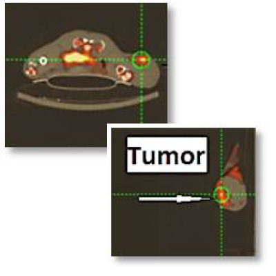
[18F]IDO49 was prepared from a tosylate precursor via two-step radiolabeling in high yields without intermediate purification. The probe has an IC50 of 95.5 ± 10.4 nM in vitro, with an IC50 of 40.9 ± 10.9 nM in a HeLa cellular assay after IDO activity was induced by IFN-ɣ exposure. [18F]IDO49 underwent no detectable degradation after 3 hours at 37ºC in PBS as measured by radio-TLC. The high binding specificity was measured by cellular uptake in HeLa cells after exposure to IFN-ɣ and a gradient of IDO inhibitor INCB24360. [18F]IDO49 accumulated selectively in tumor cells at a 2.29 ± 0.05 tumor/muscle ratio in mouse xenograft HeLa cells measured by PET 60 min after IFN-ɣ injection to confirm IDO labeling.
PUBLICATION/PATENT
- A provisional patent application was filed for Dr. Tian on August 24, 2016.
The Innovation Office
InnovationMarketing@Moffitt.org
(813) 745-6828